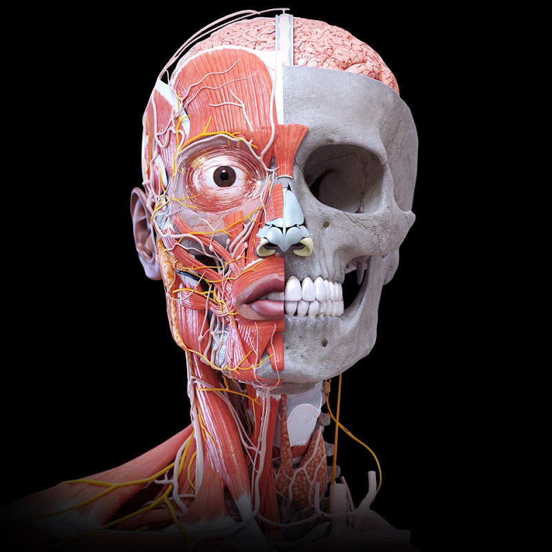
Mitral, Aortic, Tricuspid & Pulmonary Valves | A Real-Time Inner View 💓
Apr 7, 2026•Channel
AI Analysis
Data from YouTube Data API v3•Updated Just now
Video Overview
Video Details
Published1 month ago
Duration0:16
Video ID1JSxJB-0Gvw
Languageen
CategoryEducation
PrivacyPublic
Made for KidsNo
Video TypeRegular Video
Performance Metrics
Views49.9K
Likes0
Comments9
Engagement Rate0.02%
Likes per 100 views0.00
Comments per 1K views0.18
Video Tags
Description
https://www.youtube.com/@DrBahaaDmour
──────────
Each heartbeat is made up of two main sounds known as S1 and S2. In this video, you'll see exactly how the S1 sound corresponds to the closing of the mitral and tricuspid valves at the start of ventricular contraction, and how the S2 sound is produced when the aortic and pulmonary valves close as the ventricles relax.
This high-precision animation is created by Dr. Bahaa to help medical students understand the exact timing and coordination of heart sounds with valve motion. It's also made easy for a general audience to grasp how the heart works. Watch and see the perfect synchronization of these sounds with the valve movements in a truly professional and educational manner.
──────────
🎥 Video created by
🩺 Dr. Bahaa N. Dmour
⚡ CEO & Founder of Smart Doctor
──────────
#Anatomy #Heart #HumanBody #3D_Anatomy #Anatomía #Anatomi #Anatomia #Education #Smartdoctor1 #Medicina #Doctor #Smart_Doctor #DrBahaaDmour #Dr_Bahaa_Dmour #aorticvalve #mitralvalv #tricuspidvalve #pulmonaryhypertension #heartanatomy #3danimation #cardiology #anatomy #heart #cardio #meded #education #3danimation #3dmodel #animation