Behind Your Breath 🫁
Mar 15, 2026•Channel
AI Analysis
Data from YouTube Data API v3•Updated Just now
Video Overview
Video Details
Published2 months ago
Duration0:16
Video IDv2Oj8kfSHBE
Languageen
CategoryEducation
PrivacyPublic
Made for KidsNo
Video TypeYouTube Short
Performance Metrics
Views6.2M
Likes0
Comments16
Engagement Rate0.00%
Likes per 100 views0.00
Comments per 1K views0.00
Video Tags
Description
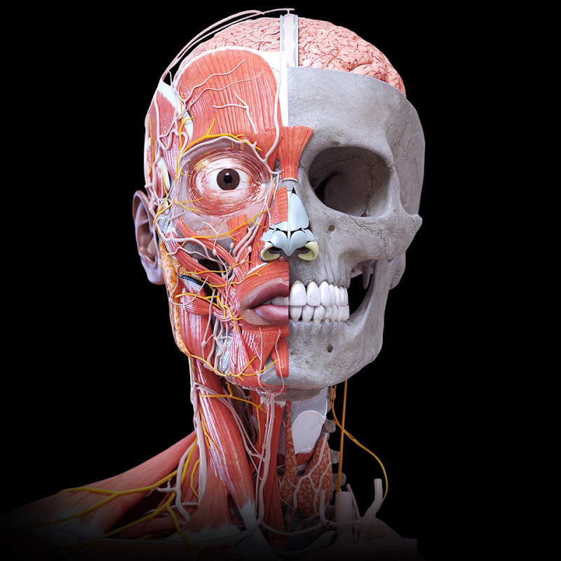
Every breath you take travels through a complex branching network called the bronchial tree. Inside the chest, the trachea divides into two main bronchi that enter the lungs and continue splitting into smaller and smaller airways until oxygen finally reaches the tiny air sacs responsible for gas exchange.
This 3D anatomical animation reveals a rare posterior view of the respiratory system, showing the relationship between the trachea, bronchi, lungs, and surrounding organs inside the thoracic cavity. From this angle you can clearly see how the airway sits in front of the esophagus while major blood vessels and nerves pass around it inside the chest.
Understanding this spatial arrangement helps explain how breathing works, why airway obstruction can affect oxygen flow, and how surgeons and physicians visualize these structures during medical procedures.
The human chest is not just a space for the lungs—it is a tightly organized anatomical chamber where airways, vessels, nerves, and organs coexist in a remarkably precise layout.-
-
🎥 Video created by 🩺 (Dr._Bahaa_N_Dmour), CEO & Founder of Smart Doctor 🪄
-
-
-
-
#Anatomy #Heart #HumanBody #3D_Anatomy #Anatomía #Anatomi #Anatomia #Education #Smartdoctor1 #Medicina #Doctor #Smart_Doctor #DrBahaaDmour #Dr_Bahaa_Dmour #Lungs #Bronchi #RespiratorySystem #HumanAnatomy #MedicalAnimation #ThoracicCavity #Anatomy3D #MedicalEducation #Biology #SmartDoctor